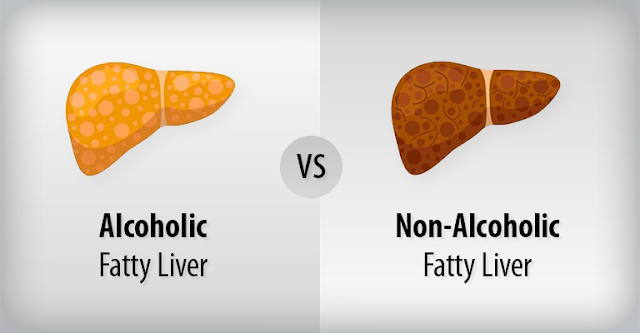
In this post, we explore the similarities and differences between alcohol-related liver disease (ARLD) and non-alcoholic fatty liver disease (NAFLD)—two of the most common causes of liver dysfunction worldwide.
Both progress through similar pathological stages but have distinct
triggers, mechanisms, and associated conditions. Let’s dive into how each
affects liver structure and function.
🔬 Shared Disease Spectrum:
Steatosis → Steatohepatitis → Fibrosis → Cirrhosis
- Both ARLD and NAFLD can follow a similar pathological progression despite differing triggers:

- Steatosis
(fatty liver): Accumulation of triglycerides within hepatocytes.
- The liver plays a central role in fat
metabolism, including storing triglycerides (TG) in hepatocytes.
- In
both ARLD and NAFLD, excess fat accumulates in liver cells, impairing
normal function.
- Alcohol (in ARLD) and insulin resistance (in NAFLD) disrupt fat breakdown and promote lipogenesis, leading to steatosis.
- Steatohepatitis:
Inflammatory cell infiltration and hepatocellular injury.
- The liver plays a central role in fat
metabolism, including storing triglycerides (TG) in hepatocytes.
- In
both ARLD and NAFLD, excess fat accumulates in liver cells, impairing
normal function.
- Alcohol
(in ARLD) and insulin resistance (in NAFLD) disrupt fat breakdown and
promote lipogenesis, leading to steatosis.
- Fibrosis
and Cirrhosis: Ongoing injury leads to scarring, regenerative nodules,
and eventually impaired hepatic function.
- Continuous injury activates hepatic
stellate cells, which deposit collagen and replace normal liver tissue
with fibrosis.
- Over
time, fibrosis disrupts liver architecture, impairing blood flow and
leading to cirrhosis—an irreversible end-stage condition.
However, despite these shared disease stages, the underlying drivers and systemic context differ.
🍺 Alcohol-Related Liver Disease (ARLD)
Pathophysiology

📌 Why does alcohol cause liver damage?
Ethanol metabolism produces toxic intermediates that disrupt liver homeostasis:
🔹 Hepatic Steatosis via NADH Overproduction
- Alcohol
is metabolized primarily by alcohol dehydrogenase (ADH) and aldehyde
dehydrogenase (ALDH) in hepatocytes.
- These
reactions increase NADH levels, which:
- 🚀 Promote lipogenesis (fat synthesis).
- 🚫 Suppress fatty acid oxidation, causing fat accumulation in hepatocytes.
🔹 Acetaldehyde Toxicity
- Acetaldehyde
(a metabolite of ethanol) is highly reactive, binding to proteins
and DNA.
- This
results in mitochondrial dysfunction, impairing hepatocyte
survival.

🔹 Oxidative Stress & Inflammation
- Ethanol metabolism through CYP2E1
produces reactive oxygen species (ROS), damaging hepatocytes.
- ROS cause lipid
peroxidation, triggering apoptosis (programmed cell death).
- Over time, chronic
inflammation leads to fibrosis and cirrhosis.
🔹 Kupffer Cell Activation & Immune Dysregulation
- Kupffer cells
(liver-resident macrophages) respond to oxidative stress by releasing TNF-α and IL-6.
- This sustained
inflammatory cascade pushes ARLD toward alcoholic hepatitis.
🔹 Microbiome Changes & Gut Permeability
- Alcohol disrupts gut
microbiota, leading to increased endotoxin absorption (via a “leaky
gut”).
- Endotoxins further trigger immune activation, worsening liver inflammation.

Clinical Features
🔬 How do these mechanisms translate into symptoms?
✔ Jaundice occurs because hepatocyte injury impairs
bilirubin conjugation/excretion.
✔
Tender hepatomegaly results from hepatocyte ballooning and inflammatory
infiltration.
✔
Ascites & hypoalbuminaemia arise due to impaired hepatic protein
synthesis and portal hypertension.
✔
Asterixis & encephalopathy occur when liver detoxification fails,
leading to ammonia accumulation.
✔
AST predominance (>2) reflects mitochondrial alcohol toxicity, as AST
is more concentrated in mitochondria than ALT.
Key
takeaway: Alcohol
metabolism directly drives hepatocyte injury via oxidative stress,
inflammation, and fibrosis. Abstinence halts this progression.

🥦 Non-Alcoholic Fatty Liver Disease (NAFLD)
Pathophysiology
📌 Why does metabolic dysfunction cause liver injury?
NAFLD arises from insulin resistance and lipid toxicity, which disrupt liver metabolism:
🔹 Insulin Resistance → Increased Free Fatty Acids
- Normally, insulin suppresses fat
breakdown (lipolysis) and regulates glucose uptake.
- In insulin resistance:
- Excess free fatty acids (FFAs) flood the liver due to uncontrolled lipolysis.
- The liver converts excess
FFAs into triglycerides, leading to hepatic steatosis.
🔹 Lipotoxicity & Mitochondrial Dysfunction
- FFAs generate reactive
oxygen species (ROS), damaging hepatocytes (similar to ARLD).
- ROS trigger lipid
peroxidation, leading to cell injury and inflammation.
- Over time, oxidative damage
causes hepatocyte apoptosis and fibrosis progression.
🔹 Gut-Liver Axis & Chronic Inflammation
- Dysbiosis in obese and
insulin-resistant individuals allows gut-derived endotoxins to
enter circulation.
- These activate Kupffer
cells, sustaining a chronic inflammatory environment that
promotes NASH progression.

Clinical Features
🔬 How do these mechanisms translate into symptoms?
✔ Hepatomegaly is common due to fat accumulation and
mild inflammation.
✔
ALT predominance ( is due to metabolic-driven hepatocyte injury
(where ALT remains higher).
✔
Silent progression occurs because fibrosis develops slowly,
unlike ARLD.
✔
Association with obesity, hypertension, and T2DM reflects NAFLD’s
systemic metabolic origins.
✔
Advanced disease can lead to non-alcoholic steatohepatitis (NASH),
fibrosis, cirrhosis, and hepatocellular carcinoma—even in non-obese individuals
(“lean NAFLD”).
Key
takeaway: NAFLD
is driven by insulin resistance and lipid toxicity rather than ethanol
exposure. Metabolic control (weight loss, glycaemic regulation) is the key to
halting disease progression.

🔍 Key Differences
Feature | ARLD | NAFLD |
Trigger | Alcohol consumption | Metabolic syndrome |
AST:ALT | >2 | <1 |
Reversibility | Improves with abstinence | Improves with weight loss/metabolic control |
Inflammation driver | Acetaldehyde, ROS, cytokines | Insulin resistance, lipotoxicity |
Comorbidities | Psychiatric illness, malnutrition | T2DM, obesity, cardiovascular risk |
Risk of HCC | Increased (esp. with cirrhosis) | Increased even without cirrhosis |
✅ ARLD and NAFLD share pathological stages but differ in
triggers.
✅ Understanding
the “HOW” explains why symptoms emerge and how disease progresses.
✅ Early
intervention can halt progression before irreversible cirrhosis develops.

Patients can display a mixed picture with both hepatic steatosis AND alcohol related liver disease so it is important to take a thorough history and examination to identify contributing factors.
🩺 Therapeutic Strategies
🍺 ARLD: Abstinence & Supportive Care
📌 Why is abstinence the cornerstone?
Alcohol directly damages hepatocytes through acetaldehyde toxicity, oxidative stress, and chronic inflammation. Unlike metabolic disease, which progresses slowly, ongoing alcohol use accelerates liver injury—making abstinence the only intervention that halts damage immediately.✅ Eliminating Alcohol → Stops ROS Generation & Inflammation
- Ethanol
metabolism via CYP2E1 generates reactive oxygen species (ROS),
worsening hepatocyte apoptosis.
- Abstinence
removes the oxidative stress burden, allowing hepatocytes to
recover before fibrosis becomes irreversible.
✅ Can the Liver Regenerate?
- Yes—early-stage
steatosis and mild fibrosis can reverse with sustained alcohol
cessation.
- However,
once cirrhosis develops, fibrosis becomes largely irreversible.
✅ Why Nutritional Support?
- Alcohol
disrupts gut absorption, leading to thiamine, folate, and
protein deficiency.
- Chronic
use causes malnutrition and muscle wasting, worsening liver
function.
- Supporting nutrient replenishment is key for hepatocyte regeneration.
💊 Medical Management Explained
🔹 Steroids (for severe alcoholic hepatitis)
- In
advanced cases, TNF-α driven inflammation leads to hepatocyte
necrosis.
- Corticosteroids
blunt the inflammatory cascade, reducing mortality in high-risk
patients (MDF >32).
- However,
infection screening is crucial—steroids impair immune function,
making infections a major risk.
🔹 Lactulose (for
encephalopathy)
- Alcohol-related
liver injury reduces ammonia clearance, leading to cognitive
dysfunction (encephalopathy).
- Lactulose
traps ammonia in the gut, preventing neurotoxic effects.

🔹 Ascites Control
(Diuretics & Salt Restriction)
- Portal
hypertension drives fluid accumulation in the peritoneal cavity.
- Reducing
sodium intake and using diuretics helps manage ascites and prevent
complications like spontaneous bacterial peritonitis.
ARLD is dose-dependent toxicity—remove
the toxin, and liver function can stabilize.

Pathophysiology of ascites
🥦 NAFLD: Metabolic Control & Fibrosis Prevention
📌 Why does weight loss help?
Unlike ARLD, NAFLD isn't driven by an external toxin—it's a consequence of metabolic dysfunction, particularly insulin resistance and lipotoxicity. Since excess fat deposition fuels hepatic inflammation, reversing metabolic dysfunction removes the source of injury.✅ Weight Loss → Reduces Fat Accumulation & Inflammation
- Losing
≥7-10% of body weight significantly reduces hepatic steatosis.
- Weight
loss also improves insulin sensitivity, lowering free fatty acid
burden before fibrosis progresses.
✅ Exercise → Enhances Insulin Sensitivity & Fat Metabolism
- Regular
physical activity reduces visceral fat stores, lowering hepatic fat
deposition.
- Exercise
modifies inflammatory pathways, reducing liver fat accumulation
and oxidative stress.
💊 Medical Management Explained 🔹 Pioglitazone (PPARγ Agonist)
- PPARγ
regulates fat metabolism and insulin sensitivity.
- Pioglitazone
reduces hepatic inflammation, slowing NASH progression.
🔹 GLP-1 Receptor
Agonists
- Initially
used for diabetes, these drugs show promise in reducing
NAFLD-related fat accumulation.
- They
enhance satiety and weight loss, indirectly reducing hepatic fat
burden.
🔹 Vitamin E (for
non-diabetic NASH)
- Acts
as an antioxidant, reducing oxidative damage to hepatocytes.
- Not
a universal treatment but useful in select cases.
🛑 Fibrosis Surveillance Explained
✅ Why is fibrosis staging
critical?
- NAFLD
can silently progress to cirrhosis, even in non-obese individuals.
- Since
patients often lack symptoms, non-invasive markers (FibroScan, NAFLD
fibrosis score) help detect advanced disease early.
✅ Why HCC screening?
- NAFLD
increases hepatocellular carcinoma (HCC) risk, even in the
absence of cirrhosis.
- Advanced
fibrosis warrants regular imaging to catch malignancy early.
NAFLD is metabolic overload—reducing
fat burden allows hepatocytes to recover before irreversible fibrosis
develops.

🔎 Clinical Case Examples
Case 1: Alcohol-Related Liver Disease (ARLD)
Presentation
A 52-year-old male with 15-year history of
alcohol use (~60g/day) presents with:
- Symptoms: Fatigue, anorexia, jaundice, RUQ
discomfort, and increasing abdominal distension.
- Systemic Features: Easy bruising, lower
limb swelling.
Examination Findings
✅ General:
- Appearance: Cachectic, mild confusion (possible
hepatic encephalopathy).
- Vitals: HR 96 bpm, BP 100/65 mmHg, Temp 37.7°C
(low-grade fever in alcoholic hepatitis).
✅ Abdominal
Exam:
- Inspection: Jaundice, spider naevi, palmar erythema.
- Palpation: Tender hepatomegaly (liver
edge firm, irregular ± nodular).
- Ascites: Shifting dullness present.
- Other Findings: Splenomegaly suggesting
portal hypertension.
✅ Systemic Exam:
- Neurological: Asterixis present
(suggesting hepatic encephalopathy).
- Dermatological: Ecchymosis and capillary
fragility (due to coagulopathy).
Investigations
|
Test |
Result |
Reference Range |
|
AST |
210 IU/L |
10–40 IU/L |
|
ALT |
90 IU/L |
10–45 IU/L |
|
AST:ALT Ratio |
>2 |
Typically <1 |
|
Total Bilirubin |
45 µmol/L |
|
|
Albumin |
30 g/L |
35–50 g/L |
|
INR |
1.8 |
0.9–1.3 |
|
GGT |
150 IU/L |
10–60 IU/L |
|
ALP |
120 IU/L |
30–110 IU/L |
Ultrasound: Coarse hepatic echotexture, splenomegaly, ascites
Clinical Reasoning:
The combination of jaundice, tender hepatomegaly, AST>ALT, coagulopathy, and ascites strongly suggests alcoholic hepatitis with cirrhotic complications.🔹 Why no FibroScan initially? Liver stiffness is unreliable in acute inflammation, so we focus on clinical severity (INR, bilirubin) and re-evaluate fibrosis once stable.
🔹 Key questions: Steroid consideration (Maddrey’s score), micronutrient repletion (thiamine, folate), alcohol withdrawal management.
Case 2: Non-Alcoholic Fatty Liver Disease (NAFLD)
Presentation:
A 45-year-old female with BMI 32, hypertension, T2DM presents with:- Symptoms: Asymptomatic but noted fatigue
and mild RUQ discomfort.
- Incidental finding: Elevated ALT on routine
bloodwork.
Examination Findings
✅ General:
- Appearance: Obese (central adiposity).
- Vitals: HR 78 bpm, BP 145/85 mmHg.
✅ Abdominal Exam:
- Inspection: No jaundice or stigmata of
chronic liver disease.
- Palpation: Liver palpable 2cm below
costal margin (suggesting steatosis).
- No ascites or splenomegaly.
✅ Systemic Exam:
- Metabolic Features: Acanthosis nigricans
(insulin resistance).
- Cardiovascular Risk: Mild ankle oedema
(suggestive of early metabolic dysfunction)
Investigations
|
Test |
Result |
Reference Range |
|
AST |
55 IU/L |
10–40 IU/L |
|
ALT |
85 IU/L |
10–45 IU/L |
|
AST:ALT Ratio |
<1 |
Typically <1 |
|
Fasting Glucose |
6.8 mmol/L |
3.9–5.8 mmol/L |
|
HbA1c |
7.2% |
<5.7% |
|
Lipid Panel |
||
|
Triglycerides |
2.5 mmol/L |
<1.7 mmol/L |
|
Total Cholesterol |
5.8 mmol/L |
<5.5 mmol/L |
|
LDL Cholesterol |
3.5 mmol/L |
<3.0 mmol/L |
|
HDL Cholesterol |
1.1 mmol/L |
>1.2 mmol/L |
- Ultrasound: Hyperechoic liver (suggestive of fatty infiltration)
- Fibroscan: Elevated stiffness
(suggestive of significant fibrosis
Clinical
Reasoning:
This case highlights NAFLD as a silent but progressive disease. Despite
the asymptomatic presentation, metabolic syndrome components (T2DM, obesity,
dyslipidaemia) drive hepatic injury. Fibroscan results indicate progression
beyond simple steatosis, raising concerns for non-alcoholic steatohepatitis
(NASH) and fibrosis.
📌 ALT-predominant transaminitis aligns with metabolic liver disease, with hepatic steatosis likely progressing towards NASH
🔹
Why a FibroScan here? Unlike ARLD, fibrosis assessment is crucial even
in subclinical NAFLD, as metabolic syndrome independently increases
cirrhosis risk.
🔹
Key questions: Lifestyle interventions (weight loss, glycaemic control),
cardiovascular risk stratification, long-term fibrosis surveillance
🧠 Clinical Takeaways
✅ Why ARLD vs NAFLD looks different on examination:
- ARLD: More systemic effects
(encephalopathy, coagulopathy, portal hypertension).
- NAFLD: Subtle findings until
advanced fibrosis, often tied to metabolic syndrome.
✅ Why investigations differ:
- ARLD: Focus on acute hepatic
inflammation and systemic decompensation.
- NAFLD: Non-invasive fibrosis
assessment critical for staging disease.
✅ How this informs management:
- ARLD: Early intervention, alcohol
cessation, nutritional support, infection screening.
- NAFLD: Weight loss, insulin
sensitivity improvement, cardiovascular risk reduction.
🔚 Wrapping It Up
Alcohol-related liver disease (ARLD) and non-alcoholic fatty liver disease (NAFLD) represent two distinct pathways to liver damage, yet both follow a common progression from fat accumulation to inflammation, fibrosis, and cirrhosis. ARLD is fueled by direct ethanol toxicity, making abstinence the most effective intervention. NAFLD, on the other hand, is driven by metabolic dysfunction, requiring lifestyle modification and metabolic control.
The challenge with both diseases is their silent progression—often asymptomatic until significant liver damage has already occurred. Timely recognition and intervention are essential to prevent irreversible cirrhosis and its complications.
The liver has a remarkable ability to heal and regenerate,
but only if action is taken early enough.
🩺 Final points to remember
✔ ARLD is a toxin-driven injury—abstinence removes the
primary insult, allowing recovery before fibrosis becomes permanent.
✔
NAFLD is a metabolic disorder—addressing obesity, insulin resistance,
and cardiovascular risk factors can revers the inflammatory cycle and halt disease progression.
✔
Both conditions can remain silent for years—early detection through
routine testing prevents late-stage complications.
✔
Intervention matters most before cirrhosis develops—once fibrosis
becomes extensive, reversal is difficult.
✔
The liver can heal—but only if action is taken in time.
- 📝 Complete the quiz on this topic →
- 📝 All posts on the gastrointestinal system →
- 📝 Structure and function of the liver →
- 📝 Signs of liver dysfunction →
- 📝 Acute viral hepatitis→
- 📝 Chronic viral hepatitis→
- 📝 Interpreting Hepatitis B serology →

No comments:
Post a Comment