Structure, Function, and Clinical Relevance
Understanding its structure and function will help explain why liver dysfunction produces such distinct clinical signs.
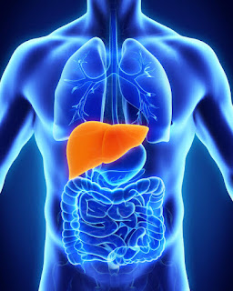
🧭 Anatomical Orientation

Macroscopic Structure
Location:
The liver sits in the right upper quadrant, beneath the diaphragm and shielded by ribs 6–10—protected from minor trauma but vulnerable in severe injuries.
Lobes:
Traditionally, the liver is divided into the right and left lobes, plus two smaller lobes—the caudate and quadrate.
Functionally, it’s divided into eight segments (Couinaud classification), each with its own vascular supply. Surgeons use this segmentation to remove diseased portions while preserving function.

Ligaments:
The falciform ligament separates the left and right lobes and anchors the liver to the anterior abdominal wall.
The ligamentum teres is a remnant of the foetal umbilical vein, which carried oxygenated blood from the placenta. After birth, it fibroses but remains a landmark in liver anatomy.

Vascular Supply:
Unlike most organs, the liver receives blood from two sources:
✅ Hepatic artery (25%) → delivers oxygen-rich blood from the systemic circulation.
✅ Portal vein (75%) → supplies nutrient-rich, oxygen-poor blood from the GI tract, pancreas, and spleen.

🩸 Why this matters:
This dual supply allows the liver to filter blood, metabolise nutrients, and detoxify toxins before circulation continues.
Blood flows through hepatic sinusoids (capillary networks) toward the central vein, draining into the hepatic veins → inferior vena cava → heart.
Sinusoids & the Space of Disse
Blood flows through hepatic sinusoids, which are low-pressure capillaries lined by fenestrated endothelial cells.

Between endothelial cells and hepatocytes lies the Space of Disse—a microscopic gap where nutrients and plasma proteins exchange freely.
Clinical relevance: In cirrhosis, fibrosis narrows the Space of Disse, reducing nutrient transfer and impairing liver function.

Microscopic Structure:
The liver is made up of hexagonal lobules, each centred around a central vein.
🟢 At each corner: A portal triad—contains branches of the hepatic artery, portal vein, and bile duct.
🔄 Blood flow: Moves from the periphery inward (portal triad → sinusoids → central vein).
🧪 Bile flow: Moves in the opposite direction—hepatocytes secrete bile into canaliculi, which drain into bile ducts.


Key Cell Types & Functions:
✅ Hepatocytes: The Workhorses of the Liver
- Functions:
- Carry out most metabolic processes, including carbohydrate, lipid, and protein metabolism.
- Synthesize plasma proteins (albumin, clotting factors, acute-phase reactants).
- Detoxify toxins and drugs via Phase I (cytochrome P450 oxidation) and Phase II (conjugation).
- Produce bile by processing cholesterol into bile acids.
- Clinical relevance:
- Hepatocyte necrosis or dysfunction leads to liver failure symptoms (jaundice, metabolic disturbances, coagulopathy).
- Fatty liver disease (steatosis) arises when hepatocytes accumulate excess lipids, disrupting normal function.

✅ Kupffer Cells: The Immune Guardians
- Functions:
- Phagocytose bacteria, dead cells, and debris from the portal blood.
- Regulate immune tolerance to gut-derived antigens.
- Secrete cytokines to influence liver inflammation and repair.
- Clinical relevance:
- In cirrhosis, Kupffer cells become overactive, triggering excessive inflammation and worsening fibrosis.
- Kupffer cell dysfunction leads to increased susceptibility to gut-derived infections (e.g., spontaneous bacterial peritonitis in ascitic patients).

✅ Stellate (Ito) Cells: The Fibrosis Architects
- Functions:
- Store vitamin A in lipid droplets when inactive.
- Mediate fibrosis during liver injury by producing extracellular matrix components (collagen).
- Regulate blood flow within sinusoids by contracting in response to injury.
- Clinical relevance:
- In chronic liver disease, stellate cells become overactivated, leading to fibrosis and cirrhosis.
- Vitamin A deficiency can result from altered stellate cell function, affecting vision and immune health.

✅ Endothelial Cells: Fenestrated Gatekeepers
- Functions:
- Line hepatic sinusoids with fenestrations (pores), allowing nutrients and plasma proteins to pass into hepatocytes.
- Regulate blood flow through the liver's sinusoids, maintaining low resistance circulation.
- Interact with Kupffer and stellate cells to mediate liver inflammation and repair.
- Clinical relevance:
- Endothelial dysfunction contributes to portal hypertension, increasing the risk of variceal bleeding in cirrhotic patients.
- Loss of fenestrations impairs nutrient exchange, affecting metabolism and protein synthesis.
✅ Cholangiocytes:
- Epithelial cells lining bile ducts, responsible for modifying bile composition by adjusting fluid and electrolytes.
- Clinical relevance: Dysfunction leads to cholestasis and biliary diseases like primary sclerosing cholangitis.
✅ Pit Cells:
- Liver-specific natural killer (NK) cells that target infected or damaged cells.
- Clinical relevance: In chronic liver disease, pit cell dysfunction contributes to increased infection susceptibility.
Hepatocyte Surface Polarity
Basolateral surface: Faces blood in the sinusoids, enabling exchange of nutrients, toxins, and proteins.
Apical surface: Faces bile canaliculi, directing bile secretion toward bile ducts.
Clinical relevance: In cholestasis (bile flow obstruction), hepatocytes retain bile pigments, leading to jaundice and liver damage.
🧠 Learning Points
💡 Why does the liver need two blood supplies?
- The hepatic artery provides oxygen to keep liver cells alive.
- The portal vein delivers nutrients & toxins for processing.
- This setup ensures the liver filters, metabolises, and detoxifies blood before it reaches the rest of the body.
💡 Why do Kupffer cells matter?
- These specialised macrophages sit within liver sinusoids and remove bacteria, toxins, and damaged cells.
- In liver dysfunction, Kupffer cells can become overactive, worsening inflammation and fibrosis
⚙️ Functions of the Liver
The liver operates like a massive biochemical factory, continuously processing, synthesising, detoxifying, and storing essential compounds. Let’s break it down into departments:

1️⃣ Metabolic Regulation: Managing Energy & Waste

Think of the liver as the body's metabolic accountant—it keeps energy levels balanced by storing and generating fuel when needed.
🟢 Carbohydrate Metabolism

✅ Glycogenesis: Stores excess glucose as glycogen for later use.
✅ Glycogenolysis & Gluconeogenesis: Releases glucose during fasting by breaking down glycogen or making new glucose from amino acids.
🔹 Mini Case:
A diabetic patient on insulin misses a meal → blood sugar drops → liver activates gluconeogenesis, producing glucose from amino acids to prevent hypoglycaemia.
🟠 Lipid Metabolism

✅ Cholesterol & lipoprotein synthesis (LDL, HDL) for cell membranes & transport.
✅ Fatty acid oxidation for energy.
✅ Lipogenesis: Converts excess carbohydrates/proteins into fat for storage.
🔹 Analogy:
The liver acts as a fat management centre—storing fat when there’s excess fuel and burning fat when energy is needed.
🔵 Protein Metabolism

✅ Transamination & deamination: Converts amino acids into usable forms.
✅ Urea cycle: Turns toxic ammonia into urea, which is safely excreted.
🔹 Mini Case:
A patient with liver failure cannot clear ammonia → it accumulates → crosses into the brain → hepatic encephalopathy (confusion, asterixis, coma).
2️⃣ Synthetic Functions: The Liver’s Production Line
The liver manufactures essential proteins that maintain circulation, clotting, and immunity.
🟢 Albumin:
✅ Maintains oncotic pressure—prevents fluid leakage from blood vessels.
✅ Low albumin → oedema & ascites (fluid accumulates in tissues)
🔹 Mini Case:
A patient with cirrhosis has low albumin → presents with swollen legs & a distended abdomen—fluid leaks from blood vessels due to reduced oncotic pressure
🟠 Clotting Factors:
✅ The liver produces fibrinogen, prothrombin, and factors V, VII, IX, X, XI for blood clotting.
✅ Liver dysfunction → bleeding risk (bruising, prolonged INR).
🔹 Analogy:
Think of clotting factors as the scaffolding holding a building together. Without enough, the structure collapses—leading to uncontrolled bleeding.
🟣 Acute-Phase Proteins: Immune Response & Inflammation
✅ C-Reactive Protein (CRP): Signals inflammation & infection.
✅ Complement proteins: Help destroy bacteria via the immune system.
🔹 Mini Case:
A patient with severe infection has a spiking fever and high CRP levels—the liver is responding to inflammation by producing acute-phase proteins.
🔵 Carrier Proteins: Transporting Essential Nutrients
✅ Transferrin: Carries iron in the bloodstream for red blood cell production.
✅ Ceruloplasmin: Transports copper, crucial for enzyme activity.
🔹 Mini Case:
A patient with iron deficiency anaemia has low transferrin levels—without enough transporter proteins, iron cannot reach the bone marrow for RBC production.
3️⃣ Detoxification: Filtering the Blood
The liver neutralises toxins, metabolises drugs, and processes bilirubin from dead red blood cells.

🟢 Drug Metabolism
✅ Phase I: Cytochrome P450 enzymes modify drugs for clearance.
✅ Phase II: Conjugation makes drugs water-soluble for excretion.
🔹 Mini Case:
A cirrhotic patient takes standard medication doses → liver fails to metabolise it properly → drugs accumulate → toxicity risk.
🟠 Bilirubin Metabolism

✅ RBC breakdown → releases unconjugated bilirubin (fat-soluble).
✅ Liver conjugates it → water-soluble bilirubin → excreted in bile.
🔹 Mini Case:
A patient with jaundice has a blocked bile duct → bilirubin backs up in blood → yellow skin & sclera + dark urine (excess bilirubin in urine).
4️⃣ Bile Production & Excretion
The liver produces bile, essential for fat digestion.
✅ Bile acids emulsify fats, aiding absorption of vitamins A, D, E, K.
✅ Bile removes bilirubin, excess cholesterol, and toxins.
🔹 Mini Case:
A patient with fat-soluble vitamin deficiency (poor bile secretion) develops easy bruising (low vitamin K), night blindness (low vitamin A).
5️⃣ Immune Surveillance: The Liver’s Defense Team
The liver helps regulate immune tolerance to gut flora & foreign antigens.
✅ Kupffer cells: Phagocytose bacteria & debris from portal blood.
✅ Stellate cells: Trigger inflammation & fibrosis if overactivated.
🔹 Mini Case:
A cirrhotic patient develops spontaneous bacterial peritonitis—weakened Kupffer cell function fails to clear gut bacteria leaking into ascitic fluid
6️⃣ Storage: The Liver’s Warehouse
✅ Glycogen → emergency glucose reserves.
✅ Iron & copper → stored for RBC production & enzyme function.
✅ Vitamins A, D, B12, K → essential for metabolism & immunity.
🧠 Takeaway for Clinical Reasoning
Think of the liver as a factory with several departments. When one department fails, it gives rise to specific signs:
• Albumin and clotting factor loss? Synthetic failure
• Confusion and asterixis? Detox failure (ammonia)
• Jaundice and dark urine? Impaired bilirubin handling
• Fatty liver and hypoglycaemia? Metabolic dysfunction
Mastering liver structure and function is key to recognising dysfunction. The liver influences metabolism, synthesis, detoxification, immunity, and storage—so its failure triggers broad systemic consequences. By understanding each functional domain, clinicians can interpret liver disease patterns with clarity, making diagnosis more intuitive.
Whether you’re linking jaundice to bilirubin metabolism, ascites to albumin synthesis, or hepatic encephalopathy to ammonia accumulation—these patterns all stem from the liver’s multitasking role. This foundational knowledge will sharpen clinical reasoning, preparing you to navigate real-world cases with confidence.
Understanding liver structure and function is the foundation for recognising dysfunction. In the next post, we'll explore how liver failure manifests—connecting physiological breakdowns to clinical signs. Stay tuned for the quiz to test your knowledge, and the next installment where we break down liver disease patterns with step-by-step reasoning

No comments:
Post a Comment